Home

congélateur dollars américain café t1 ct scan Cadre fragment Escalier

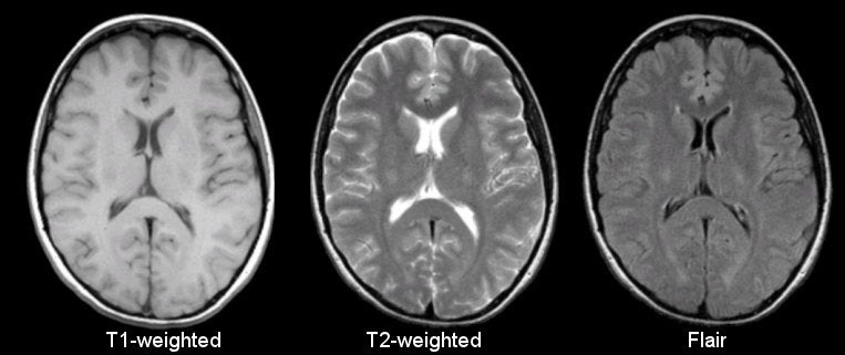
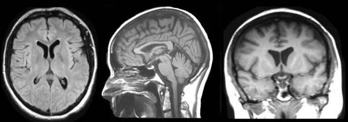
MRI Basics
MRI Basics

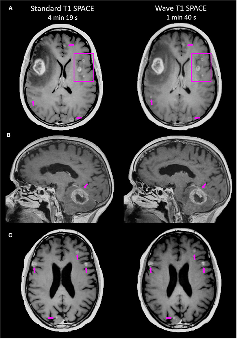
Frontiers | Accelerated Post-contrast Wave-CAIPI T1 SPACE Achieves  Equivalent Diagnostic Performance Compared With Standard T1 SPACE for the  Detection of Brain Metastases in Clinical 3T MRI | Neurology
Frontiers | Accelerated Post-contrast Wave-CAIPI T1 SPACE Achieves Equivalent Diagnostic Performance Compared With Standard T1 SPACE for the Detection of Brain Metastases in Clinical 3T MRI | Neurology

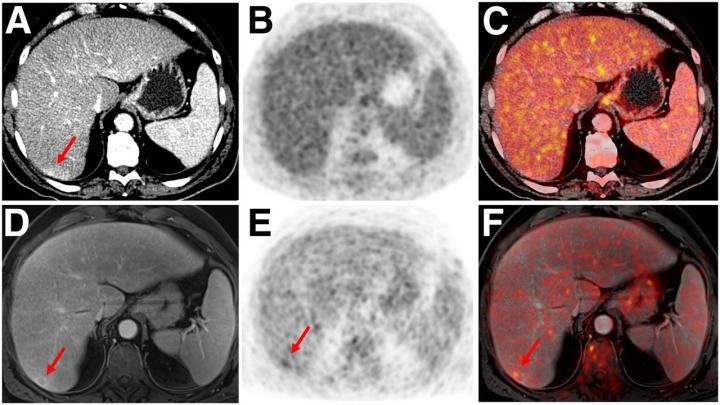
PET/MRI Outperforms PET/CT in Cancer Imaging

MRI Basics
MRI Basics

Imaging of Lung (Sagittal T1 and CE CT) - Netter Medical Artwork | Medical  artwork, Medical, Radiologic technology
Imaging of Lung (Sagittal T1 and CE CT) - Netter Medical Artwork | Medical artwork, Medical, Radiologic technology

Adrenal Tumor Classification on T1 and T2-weighted Abdominal MR Images |  Semantic Scholar
Adrenal Tumor Classification on T1 and T2-weighted Abdominal MR Images | Semantic Scholar

A Computed tomography (CT) scan image of the head. B T1-weighted image... |  Download Scientific Diagram
A Computed tomography (CT) scan image of the head. B T1-weighted image... | Download Scientific Diagram

MRI Highly Accelerated Wave‐CAIPI T1‐SPACE versus Standard T1‐SPACE to  detect brain gadolinium‐enhancing lesions at 3T - Goncalves Filho - 2021 -  Journal of Neuroimaging - Wiley Online Library
MRI Highly Accelerated Wave‐CAIPI T1‐SPACE versus Standard T1‐SPACE to detect brain gadolinium‐enhancing lesions at 3T - Goncalves Filho - 2021 - Journal of Neuroimaging - Wiley Online Library
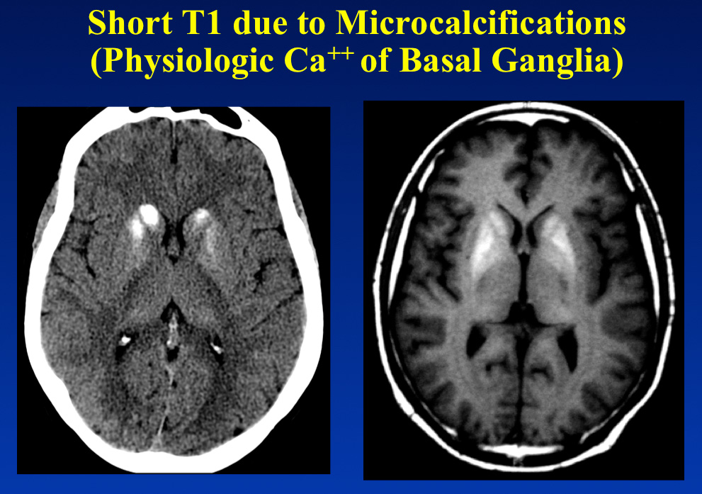
Short T1 calcification - Questions and Answers ​in MRI

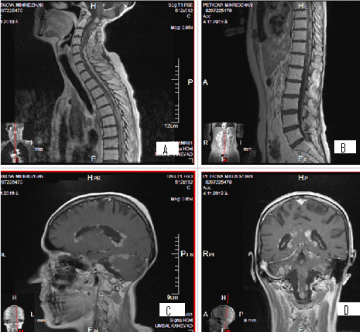
Importance of magnetic resonance imaging for diagnostics and complex  treatment in patients with Leptomeningial Disease- Six clinical cases with  a literature overview
Importance of magnetic resonance imaging for diagnostics and complex treatment in patients with Leptomeningial Disease- Six clinical cases with a literature overview
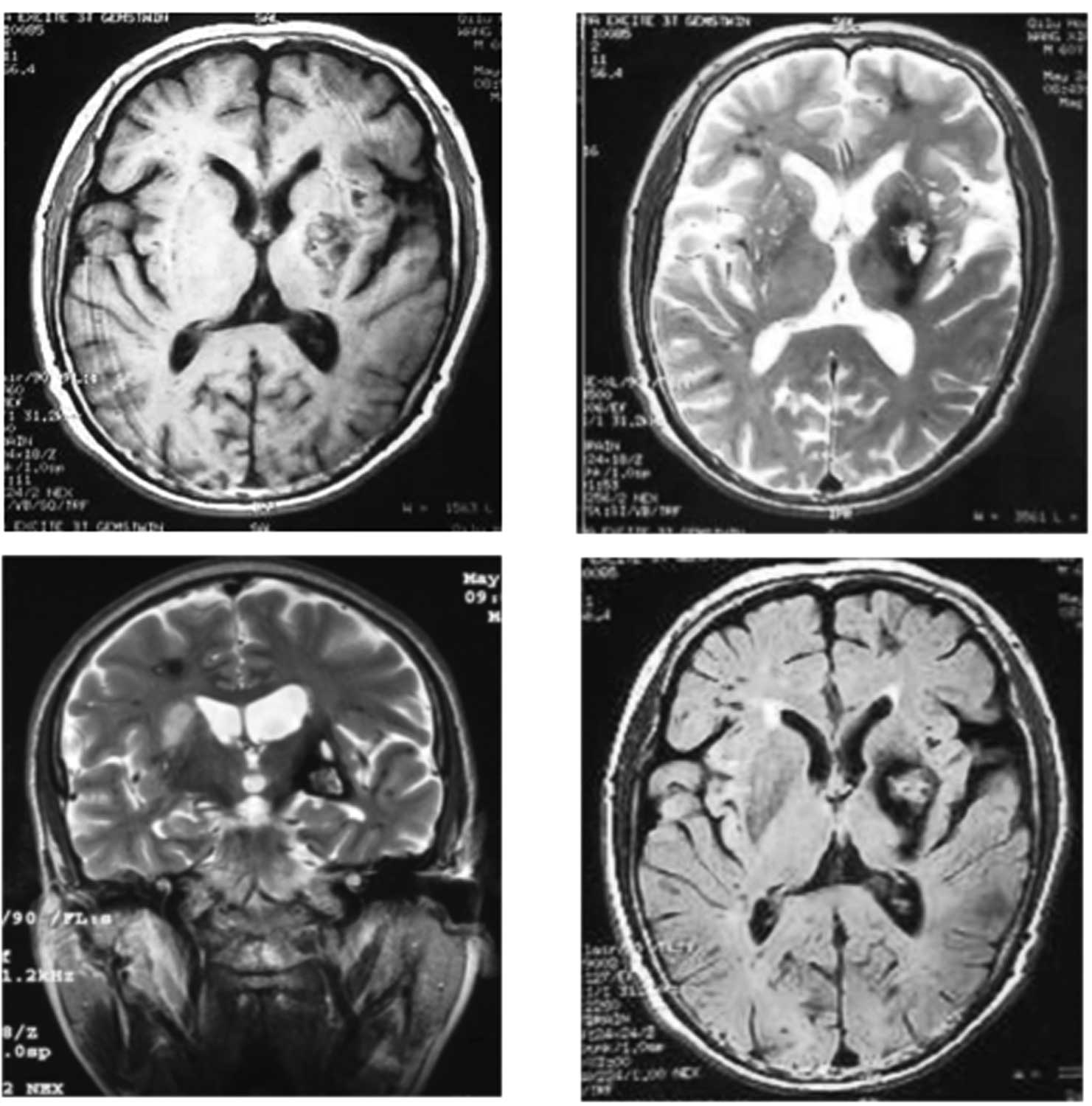
The value of T2*-weighted gradient echo imaging for detection of familial  cerebral cavernous malformation: A study of two families
The value of T2*-weighted gradient echo imaging for detection of familial cerebral cavernous malformation: A study of two families

Figure 1. [CT and T1- and T2-weighted...]. - GeneReviews® - NCBI Bookshelf

CT Scan cervical spine 3 D render . Stock Photo by ©Richmanphoto 311581240

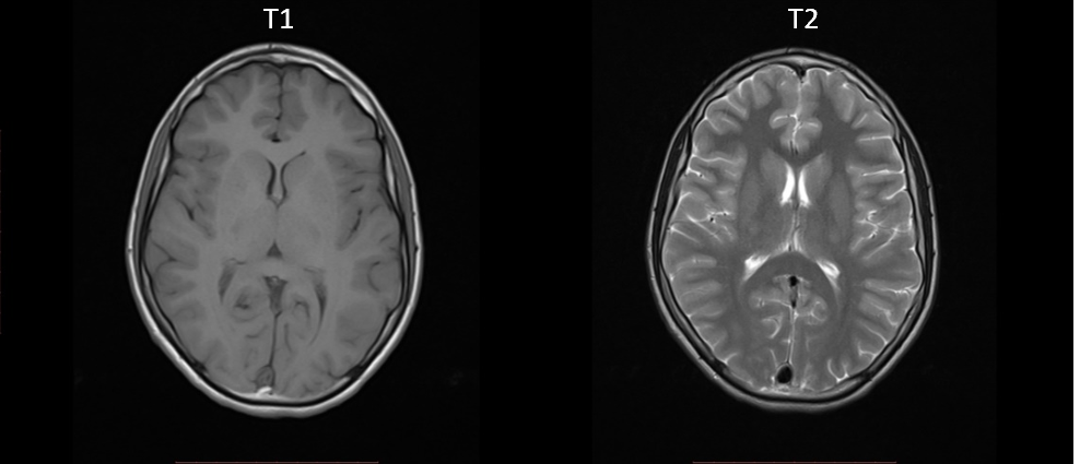
MRI brain Axial T1 and T2. stock image. Image of embolism - 154454027

CT images and T1-weighted MR images of the same patient illustrate... |  Download Scientific Diagram
CT images and T1-weighted MR images of the same patient illustrate... | Download Scientific Diagram

CERMEP-IDB-MRXFDG: A database of 37 normal adult human brain [18F]FDG PET, T1  and FLAIR MRI, and CT images available for research | bioRxiv
CERMEP-IDB-MRXFDG: A database of 37 normal adult human brain [18F]FDG PET, T1 and FLAIR MRI, and CT images available for research | bioRxiv

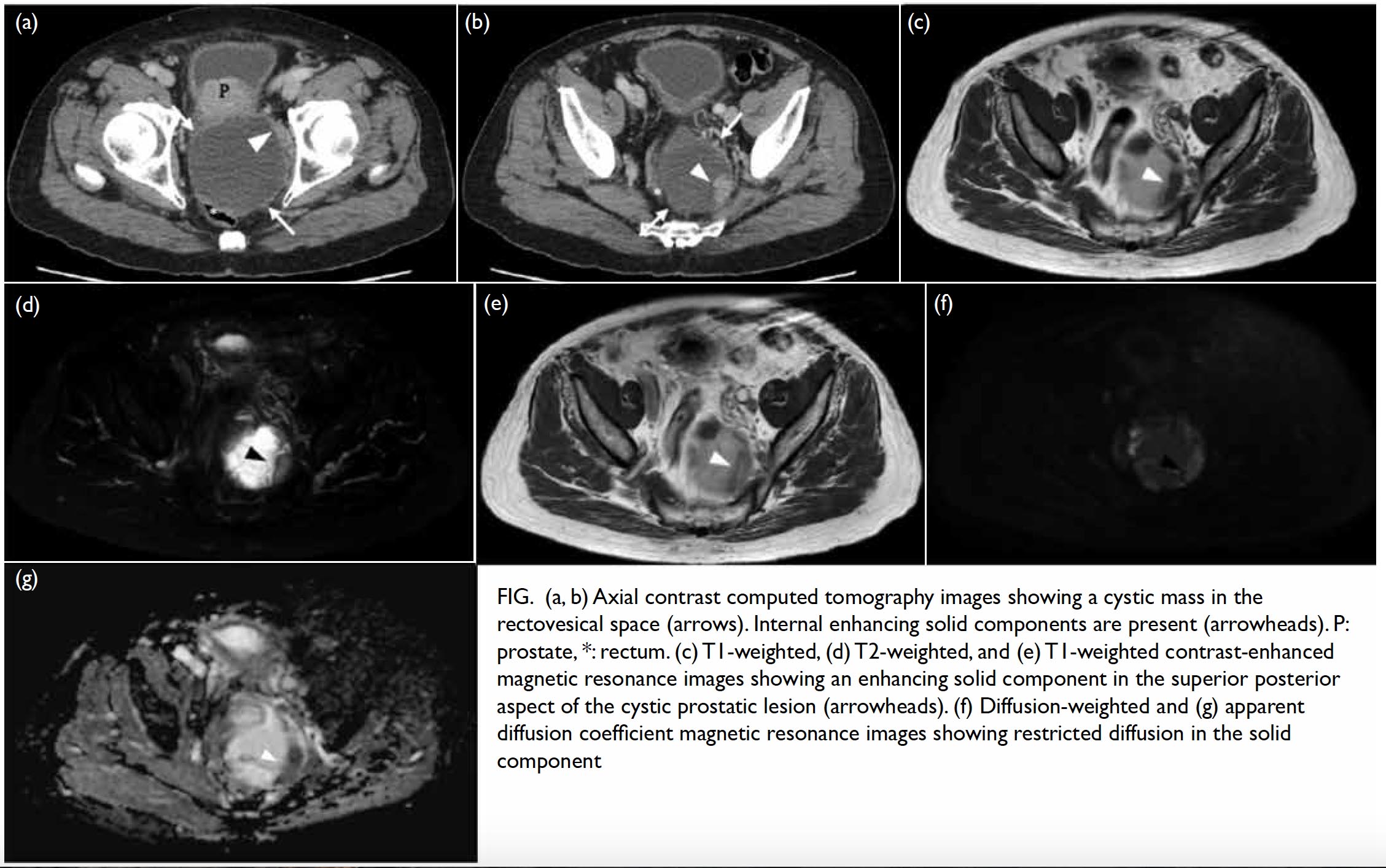
Cystic prostatic carcinoma: case report and literature review | HKMJ

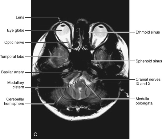
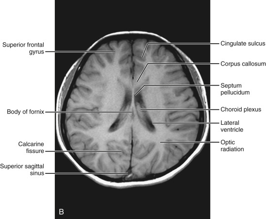
Normal Anatomy | Radiology Key

The Basics of MRI Interpretation | Radiology | Geeky Medics

Normal Anatomy | Radiology Key

PLOS ONE: 18F-Fluorothymidine PET-CT for Resected Malignant Gliomas before  Radiotherapy: Tumor Extent according to Proliferative Activity Compared  with MRI
PLOS ONE: 18F-Fluorothymidine PET-CT for Resected Malignant Gliomas before Radiotherapy: Tumor Extent according to Proliferative Activity Compared with MRI

Fig A midline Post-contrast Sagittal T1 Weighted MRI - ppt video online  download
Fig A midline Post-contrast Sagittal T1 Weighted MRI - ppt video online download

Initial CT scan with left insular hematoma. b–d MRI-T1, T2, and T1... |  Download Scientific Diagram
Initial CT scan with left insular hematoma. b–d MRI-T1, T2, and T1... | Download Scientific Diagram

Deep Learning with Magnetic Resonance and Computed Tomography Images | by  Jacob Reinhold | Towards Data Science
Deep Learning with Magnetic Resonance and Computed Tomography Images | by Jacob Reinhold | Towards Data Science

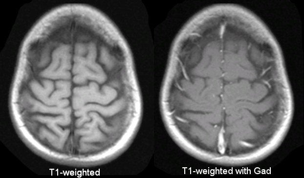
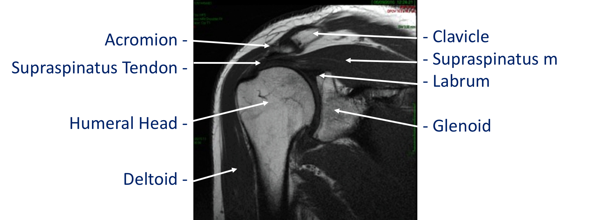
Imaging - Cambridge Shoulder

Coronal CT scan (A) and coronal contrast-enhanced T1-weighted MRI scan... |  Download Scientific Diagram
Coronal CT scan (A) and coronal contrast-enhanced T1-weighted MRI scan... | Download Scientific Diagram

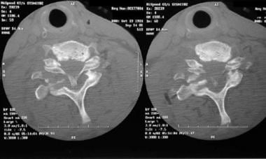
Lower Cervical Spine Fractures and Dislocations Workup: Laboratory Studies,  Imaging Studies
Lower Cervical Spine Fractures and Dislocations Workup: Laboratory Studies, Imaging Studies

MRI Basics
MRI Basics